CTSA Logomark in front of red decorative line

CTSA Wellness Guide

Understanding Your Heart and Common Heart Conditions

With our Wellness Guide, you will learn how the heart works and the important role played by the coronary arteries, valves and electrical system of the heart. You also will receive some basic information about irregular heartbeats, coronary artery disease, angina (chest pain) and heart attacks.

How the Heart Works

The heart is a muscle about the size of an adult male fist. Its job is to pump blood to all parts of the body.

The heart is made up of four chambers that hold and pump blood. The top two chambers are the right atrium and left atrium. The atria collect blood returning to the heart from the veins. The atria then release the blood into the bottom chambers through valves. Heart valves act as one-way doors to separate the chambers and keep the blood moving forward.

The bottom two chambers are called the right ventricle and left ventricle. A wall (septum) separates the two right chambers from the two left chambers. Because of this, you may hear your heart described as two pumps, the “right heart” and the “left heart.” When the ventricles contract, they force blood out of the heart to different parts of the body.

The heart is surrounded by a tough protective sac called the pericardium.

Valve Conditions

There are four heart valves that act as one way doors to keep blood moving in the right direction through the heart. Valve disease occurs when a valve doesn’t work the way it should. If a valve doesn’t close all the way, blood may leak backward. This is called regurgitation or insufficiency. If a valve doesn’t open all the way, less blood moves through the opening. This is called stenosis. Valve problems cause the heart to work harder to pump the same amount of blood. Fluid may back up in the lungs or body and can cause swelling, shortness of breath and dizziness.

Septal Defects

The atrial septum is a wall of tissue that separates the upper chambers of the heart. An atrial septal defect (ASD) is a hole or opening in this wall that fails to close during the body’s development before birth. The ventricular septum divides the two lower chambers (ventricles) of the heart. A ventricular septal defect (VSD) is an opening that can be caused by a birth defect or by an extensive heart attack.

If the hole in the septum is large enough to cause problems in pressure between two heart chambers, surgery may be needed. During surgery, the hole is covered by a patch or simply closed by sewing the edges of the opening together. Some holes particularly ASDs, can be closed with a plug that is delivered by a catheter.

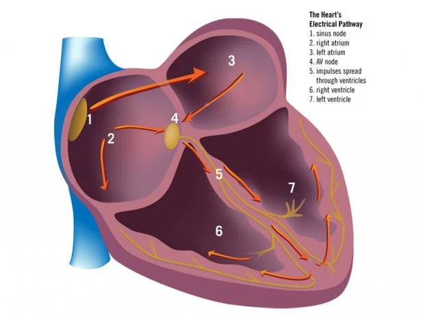
The heart’s pumping action is controlled by an electrical system that sends out impulses, or signals.

An electrical signal starts in the right atrium in the sinus node. The sinus node tells the top of the heart (atria) when and how fast to pump. It is the “pacemaker” of the heart. The signal then is sent to the AV node in the middle of the heart. The AV node holds the signal until the bottom chambers of the heart (ventricles) are filled with blood. It then sends the signal to the ventricles and tells them when to pump the blood out to the body.

Irregular Heartbeat (Cardiac Arrhythmia)

Irregular heartbeat, or cardiac arrhythmia, is a problem with your heart rhythm. The heart may beat too fast or too slowly, or may skip beats. You may feel palpitations: a rapid heart rate, skipped beats, thumping or pounding in your chest. Cardiac arrhythmias may make you feel anxious, nervous, dizzy, faint or short of breath.

Cardiac arrhythmias often are controlled with medications. Your doctor may want to do tests, however, to determine other treatments.

Premature Ventricular Beats (PVCs)

Premature ventricular beats, or PVCs, is a problem where the heart has extra beats. PVCs can occur after a heart attack or may have no known cause.

PVCs occur in the ventricles, the lower chambers of the heart, and may feel like skipped beats or extra beats. After a heart attack, PVCs may stop after the heart begins to heal. If the PVCs continue or are causing problems, your doctor may prescribe medication.

Ventricular Fibrillation (V-fib)

Ventricular fibrillation, or V-fib, is a lifethreatening arrhythmia. It comes on suddenly with chest pressure, shortness of breath and fainting. It also is known as “sudden death,” because it is often fatal. V-fib requires rapid defibrillation or shock treatment within minutes to restore a normal heart rhythm. You may have noticed automatic external defibrillators (AED) in public places. They are used to administer shock treatment. If you are at risk for V-fib, your doctor may recommend an internal cardiac defibrillator (ICD) and/or medication.

Atrial Fibrillation (A-fib)

Atrial fibrillation, or A-fib, is a problem with your heart rhythm. Symptoms of A-fib include palpitations, weakness, tiredness, shortness of breath, chest pain or tightness, dizziness or fainting.

A-fib is rarely life-threatening. It can occur following a heart attack or heart surgery, with high blood pressure or thyroid problems, or it may have no known cause. A-fib is very common, affecting people of all ages. It can lead to other problems such as stroke or heart failure. However, these problems can be avoided if the condition is managed.

A-fib occurs in the upper chambers (atria) of the heart. The cells in the atria send out extra signals, making the atria beat very quickly and unevenly. Sometimes the atria beat so fast that they may quiver instead of contracting. If the atria don’t contract, they can’t move enough blood into the ventricles. Blood that isn’t moving can easily form into clots. These clots can then move to other parts of the body causing serious health problems such as stroke.

Your doctor may prescribe tests that will help diagnose your A-fib. These tests may include EKG, echocardiogram or electrophysiology studies (EPS).

A-fib often is controlled with medications. The three most common medications that are used to slow down the speed of the heart rate are digoxin, calcium channel blockers and beta blockers. You also may be given medication to prevent clots from forming in the atria. These medications are called anticoagulants. The most common anticoagulant is called Coumadin® (warfarin). When medications don’t work sometimes the abnormal tissue is ablated or destroyed with heat or cold. This can be done with a catheter or doing heart surgery.

The coronary arteries lie on the outside surface of the heart.

The right coronary artery (RCA) supplies blood to the right side of the heart. The left coronary artery starts as the left main artery and divides into the left anterior descending (LAD) and circumflex (Cx) arteries. The left coronary artery supplies blood to the left side of the heart. Coronary arteries are about the size of cooked spaghetti. Many smaller branches lead from these main arteries.

The heart gets its blood supply from the coronary arteries. Blood is pumped from the left ventricle into the aorta, a large artery coming off of the left ventricle. The aorta supplies blood to the arteries of the heart as well as to the rest of the body.

Problems occur if the left ventricle is damaged and cannot pump enough blood to the coronary arteries. If blockages in the coronary arteries prevent blood from flowing smoothly, the heart muscle may not receive enough oxygen and nutrients to pump blood as it should.

Coronary Artery Disease

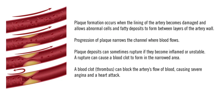
The inside of a coronary artery has a smooth lining through which blood easily flows. Coronary artery disease starts when the lining of the coronary artery is damaged. This often is due to a risk factor, such as smoking, hypertension or diabetes. A plaque deposit (a fatty material composed of cholesterol and other abnormal cells) forms between layers of the artery wall. This buildup, called atherosclerosis, causes the arteries to become stiff and slows the flow of blood to the heart muscle. Over time, buildup of stable plaque can clog the artery and cause a partial or total blockage.

Another way a coronary artery blockage can occur is when a plaque deposit becomes inflamed and unstable and actually ruptures inside the artery (see figure below). A rupture can narrow the artery even more and can cause a blood clot to form. If the blood clot is large enough, it will cut off blood flow immediately, and severe chest pain or a heart attack will result. If the heart muscle does not receive enough blood and oxygen, it is called ischemia. A symptom of ischemia is chest pain, called angina.

diagrams showing plaque formation and progression through arteries

Angina is not a disease but is a symptom of heart trouble. It is a pain or discomfort that happens when part of the heart muscle is not getting enough blood. It is usually brief, lasting just a few minutes.

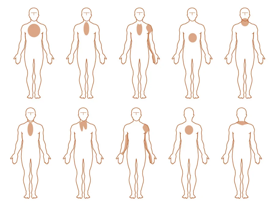
You may have pressure, tightness or pain in your chest, arms, shoulder, neck, jaw or upper back, or have stomach indigestion.

Angina discomfort often is temporary. It usually occurs during physical activity, after eating a heavy meal, after being outside when the temperature is below 32° F or above 80° F or during emotional situations.

When angina occurs:

  • Stop the activity. This is a sign your heart is not getting enough oxygen.
  • Rest.
  • If the pain does not go away in one to two minutes, place a nitroglycerin tablet under your tongue and sit down.
  • If the pain does not go away in five minutes, take another nitroglycerin.
  • If the pain still doesn’t go away, call 911, take another nitroglycerin and one uncoated aspirin. The aspirin acts as a blood thinner.

Ways to avoid angina include:

  • Eat small, frequent meals.
  • Avoid heavy exercise or physical activity for one hour after eating.
  • Check your heart rate during activity and do not exceed 20 beats above resting.
  • Do not stay out for long periods in very hot or very cold temperatures (above 80° F, below 32° F or high humidity).
  • Avoid hot tubs and saunas.
  • Manage stress.

WARNING SIGNS

Angina

Symptoms may occur with or without activity and do not stop with rest.

Chest tightness, pressure, burning – with or without pain – may go to arms or back.

Shortness of breath, extreme tiredness, dizziness, nausea, sweating, heartburn.

Heart Attack

Symptoms may occur with or without activity and do not stop with rest.

Chest tightness, pressure, burning – with or without pain – may go to arms or back.

Shortness of breath, extreme tiredness, dizziness, nausea, sweating, heartburn.

Where angina discomfort can occur:

diagram depicting various locations on the human body angina discomfort can occur

A heart attack occurs when a blood vessel (coronary artery) that feeds the heart becomes blocked and cuts off blood flow to the heart muscle (myocardium).

This blockage can occur from either fatty deposits (plaque), a spasm in the blood vessel or a clot.

A heart attack damages the heart muscle. The damage may be slight or extensive, depending on which blood vessel was blocked and how long the blood flow was cut off to the muscle. Observance of certain lab results and changes in your EKG over 24 to 48 hours will diagnose a heart attack.

Your doctor may recommend additional lab work and cardiac testing to determine if further treatment is needed. Your doctor also will prescribe medicines to help reduce your heart’s workload, to help the arteries heal and to improve blood flow. Most patients will have a cardiac catheterization to determine which artery is blocked and have added procedures such as angioplasty, a stent placement or possibly surgery.

During your hospital stay, your health team will monitor you closely, watching for any complications. If they occur, it is usually within a few days after a heart attack.

Common complications are:

  • Heart rhythm problems
  • Heart pumping problems or heart failure
  • Inflammation or swelling around the heart (pericarditis)

The heart muscle heals slowly. Scar tissue forms where the heart muscle fibers were damaged and this new tissue may not contract as well as the original tissue. The healthy heart muscle near the scarred area has to work harder and, over time, tiny new blood vessels grow from the nearby arteries to carry blood to and around the damaged muscle. These little “natural bypasses” are called collateral circulation.

Before going home, you and your loved ones will receive detailed information about your medications, your cardiac risk factors, your activity progression at home. You will be instructed when to follow up with your doctor and begin the outpatient cardiac rehabilitation program (see “Lifestyle Changes for a Healthier Heart” section).

Managing Your Care at Home

The healing process has begun. Going home is a welcoming thought, but fears and concerns can accompany it. This is normal. After all, you were monitored closely for several days at the hospital and now you will be managing on your own. Your goals for the first two weeks are to follow the going home instructions, walk daily as prescribed and relax. Remember, your body is adjusting to a new health condition and to new medications that may make you feel more tired than usual.

Sit down, put your feet up and take a nap if you are tired. Listen to your body. This is the time to let others help you. Do activities that are easy and enjoyable such as playing games, watching movies, reading and just talking.

Most people do not return to work until after they have a follow-up visit with the doctor and start a cardiac rehabilitation program. Use your progressive walking program (see bottom of page) to guide you with your daily walking and refer to the “Exercising for a Healthier Heart” section in this binder for further guidelines. Remember, this lower level of activity is temporary.

It is common for you (and your partner) to feel a wide range of emotions after experiencing a heart attack. Limiting your activities, being out of your normal routine and becoming bored can affect the way you feel. At times, you may find yourself feeling tearful and depressed or you may feel overwhelmed with thankfulness and gratitude. Other common feelings are anger with yourself and those closest to you, discouragement when you don’t think you are improving as fast as you should or just being quietly scared that it will happen again. Express your feelings. Sharing with others can make the road to recovery a lot less bumpy.

Returning to sexual activity may be a concern of yours. You may not feel any desire for sexual activity at first, or you may worry it will trigger another heart attack. Actually, your risk for another heart attack during sex is low. Ask your doctor when you can return to sexual activity. In most cases, you can resume when you are able to climb two flights of stairs without chest pain, shortness of breath or an irregular heart rate. Meanwhile, showing affection with hugs, caresses and kisses is a good way to get back in touch with your partner.

Having a heart attack is a life-changing event. It is a time to become aware of what risk factors caused the problem and to make appropriate lifestyle changes (see section on “Lifestyle Changes for a Healthier Heart”).

Your doctors and a cardiac rehabilitation program will guide you on this new journey to wellness.
Progressive Walking Program

Your nurse educator will explain a walking program to you before you leave the hospital. You should begin your walking program the day after you come home from the hospital. Use the walking schedule below to chart your progress. If you are walking outdoors avoid extreme temperatures and walk on level ground, avoiding hills and grades. Please refer to the section on “Exercising for a Healthier Heart” for more guidelines.

WALKING SCHEDULE

Dates     Time

  • 3 to 5 minutes, ________ times a day
  • 6 to 10 minutes, _______ times a day
  • 12 to 15 minutes, ______ times a day
  • 18 to 20 minutes, ______ times a day
  • 25 minutes, once a day
  • 30 minutes, once a day